What is Atomic Force Microscopy
April 24, 2012
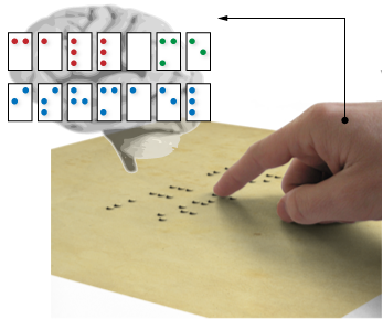
Imagine the act of reading a page of braille. Raised dots on the page are felt by a finger as it moves along the page. As the finger passes over the bumps, the forces between the paper and the finger trigger the nerve cells in the fingertip and the mind generates a spatial mapping of these forces, which is really just the arrangement of these dots.
With enough practice and training, the finger can quickly pass over each dot, and the person can read the dots.
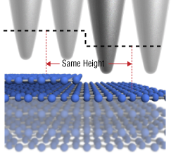
Let’s take this idea, and shrink it down in size. So, instead of millimeter size bumps on a piece of paper, let’s try and feel something much smaller, like atoms. Clearly, our finger will be too big and clumsy for this operation. We’ll need something that’s about as small as the atoms themselves. Something like a very sharp needle might work.
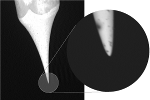
Here is tungsten wire, that’s been chemically etched to make the end terminate in a very sharp tip. While it’s not quite as small as an atom, it’s getting pretty close to that big.
Now we need some atoms to try and feel. Fortunately, there are some materials that have a really well-defined surface structure at that atomic level. Something like graphite for example. Graphite is made of carbon, and each carbon atom is stuck in a very specific place, a certain distance away from each of its neighbors. This creates a crystal lattice.
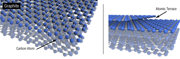
Planes of carbon atoms stacked together form the standard graphite mineral. The layers or can start and stop, leading to steps along the surface of the graphite. These terraces are exactly the height of the interlayer spacing layer: .35 nanometers
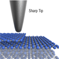
Now, we bring our finger, or in this case, the ultra-sharp metal probe near the surface, very carefully making sure it doesn’t get too close. At this point, the tip is just a few nanometers away from the surface of the graphite. Now, a few interesting things happen. Because the tip is so close, it begins to interact with the surface.
This interaction is really the heart of the AFM. Since there are numerous interactions that can occur between the tip and the sample, there exist many modes of operation. The standard method of force microscopy involves monitoring forces that depend on how far away the tip is from the sample.
By tracking these forces, we can keep the tip hovering exactly a certain distance above the surface. It’s easy to imagine moving the tip back and forth over an area, and recording the information about the position of the tip.
This is the part of the afm that requires a lot of instrumentation expertise and a very patient operator. It’s not easy keeping a tiny tip hovering just a few nanometers away from the surface. If something goes wrong during scanning, the tip can crash into the surface. This is undesirable.
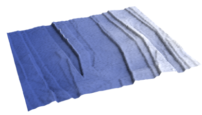
This image is a topographic scan of some terraces on graphite. It took about 5 minutes for the tip to scan over the whole area (it’s about 3 micrometers square).
This is just the beginning of what we can do with force microscopes. As mentioned above, there are many forces at play between the tip and the surface, and by playing with them all, we can probe many different characteristics of matter.
Share on: